
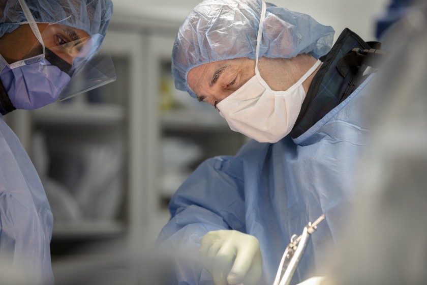
Precision is critical in spinal fracture surgery. Even small errors in hardware placement can compromise stability, injure nerves or reduce long-term outcomes. To reduce these risks, surgeons rely heavily on intraoperative imaging technologies that provide real-time visualization of anatomy. Dr. Larry Davidson, a renowned neurosurgeon specializing in spinal care, has emphasized that tools, such as the O-arm and fluoroscopy, are central to improving both safety and accuracy during complex procedures.
Integrating these imaging systems into surgical workflows, physicians can confirm hardware placement, adjust techniques immediately and make sure that fractures are stabilized, effectively. Intraoperative imaging represents a vital partnership between technology and surgical expertise, one that continues to refine outcomes for patients facing spinal injuries.
Why Intraoperative Imaging Matters
Spinal fracture surgeries often involve placing screws, rods or cages in fragile or distorted anatomy. Without imaging, the risk of misplacement is significant. Malpositioned hardware can lead to nerve compression, vascular injury or instability that requires revision.
Intraoperative imaging addresses these challenges by allowing surgeons to see exactly where instruments and implants are located, relative to the spine. It reduces guesswork and increases confidence in achieving alignment and stability. For patients, it translates into safer surgeries and fewer complications.
Fluoroscopy: Real-Time Visualization
Fluoroscopy has long been a cornerstone of spinal surgery. Using continuous X-ray technology, it provides real-time images of the spine as surgeons place screws or perform decompressions. In fracture surgery, fluoroscopy allows confirmation of needle trajectories, screw placement and implant alignment.
Its mobility makes it useful in a variety of settings, from minimally invasive procedures to complex reconstructions. While fluoroscopy exposes both patients and staff to radiation, advances in technique and shielding have reduced risks. Surgeons trained in efficient imaging use can minimize exposure, while maintaining accuracy.
The O-arm: Three-Dimensional Precision
The O-arm represents a more advanced form of intraoperative imaging. Unlike traditional fluoroscopy, it provides three-dimensional images of the spine, similar to a CT scan, during the operation. These high-resolution images allow for precise planning and real-time adjustments.
The O-arm is especially valuable when anatomy is distorted or hardware placement is complex, providing detailed cross-sectional views that clarify depth, angle and trajectory, while reducing the risk of misplacement. When paired with navigation systems, it guides instruments with GPS-like precision. Dr. Larry Davidson notes that this technology has transformed his ability to manage difficult fractures, particularly in patients with osteoporosis or multiple injuries, offering greater precision, confidence and a lower likelihood of revision surgery.
Combining Fluoroscopy and O-arm
Many surgical centers use fluoroscopy and O-arm imaging in combination. Fluoroscopy provides continuous guidance during hardware insertion, while the O-arm delivers confirmation scans that verify placement. This dual approach maximizes safety, while balancing efficiency. For example, surgeons may use fluoroscopy to guide screw trajectories, and then perform an O-arm scan to confirm that placement is accurate. If adjustments are needed, they can be made immediately, avoiding complications later.
Benefits for Patients
The benefits of intraoperative imaging extend directly to patients. Accurate hardware placement reduces the risks of nerve injury, blood loss and instability. Better alignment improves outcomes, including pain reduction and mobility restoration. Intraoperative confirmation also lowers the need for revision surgeries, which are more complex and carry higher risks. For patients, this means smoother recoveries and greater confidence in the durability of their treatment.
Dr. Larry Davidson explains, “Combining advanced robotic tools with surgical expertise elevates patient care, by making procedures safer and recovery more manageable.” Intraoperative imaging embodies this principle, reinforcing how technology enhances precision, while allowing surgeons to focus on protecting function and supporting long-term stability.
Risks and Limitations
Despite its advantages, intraoperative imaging carries considerations. Radiation exposure, though reduced with modern techniques, remains a factor. O-arm systems also require significant resources, including specialized equipment and trained staff. Not all hospitals or surgical centers have access to O-arm technology, limiting its availability. Cost can also influence whether patients are offered this level of precision. Surgeons must balance these factors, while making sure that patients still receive safe, effective care.
Athletes and Intraoperative Imaging
Athletes undergoing fracture surgery can gain significant advantages from intraoperative imaging. Their spines face high demands during recovery and competition, making precise hardware placement critical. Imaging helps align surgical outcomes with performance goals, while minimizing the risk of complications that could impact their careers.
For athletes, the combination of fluoroscopy and O-arm provides reassurance that surgical corrections are stable enough to withstand future demands. Rehabilitation can then focus on rebuilding strength and performance with confidence in the surgical repair.
Training Surgeons in Imaging Techniques
Incorporating intraoperative imaging into surgical practice requires training. Surgeons must learn to interpret images quickly, minimize radiation exposure and integrate imaging with navigation systems. Simulation labs provide opportunities to practice these skills in controlled environments.
Training is just as important as the technology. Even the most advanced imaging can’t guarantee safety, without skilled interpretation. Mentorship and hands-on experience are essential for guiding younger surgeons in using these tools, effectively and responsibly.
Advances on the Horizon
Research continues to refine intraoperative imaging. Low-dose radiation systems, improved navigation and AI-assisted interpretation promise to make imaging safer and more efficient. Portable CT scanners may expand access, bringing advanced imaging to more hospitals and patients. Integration with robotics represents another frontier. By combining real-time imaging with robotic precision, surgeons may achieve levels of accuracy that further reduce complications and speed recovery.
Patients as Partners in Care
Patients are also an important part of the imaging process. Learning how intraoperative imaging enhances safety helps them understand its value. Education on the benefits and risks of radiation supports informed consent. When patients recognize that imaging improves accuracy and lowers the chance of revision surgery, they become more engaged in their care, and more confident in their surgical plan.
The role of intraoperative imaging in fracture surgery will only expand as technology advances. Systems like the O-arm and fluoroscopy will continue to develop, offering higher precision with lower risks. As these tools become more widely available, more patients will benefit from the safety and accuracy they provide.
Intraoperative imaging has become indispensable in spinal fracture surgery, providing real-time visualization and three-dimensional precision that improve outcomes. Fluoroscopy offers continuous guidance, while the O-arm provides confirmation and accuracy, unmatched by older systems. The experience highlights the importance of combining technology with expertise. This approach shows that intraoperative imaging goes beyond hardware placement, supporting safety, stability and confidence in every fracture repair.